Introduction to Nevus
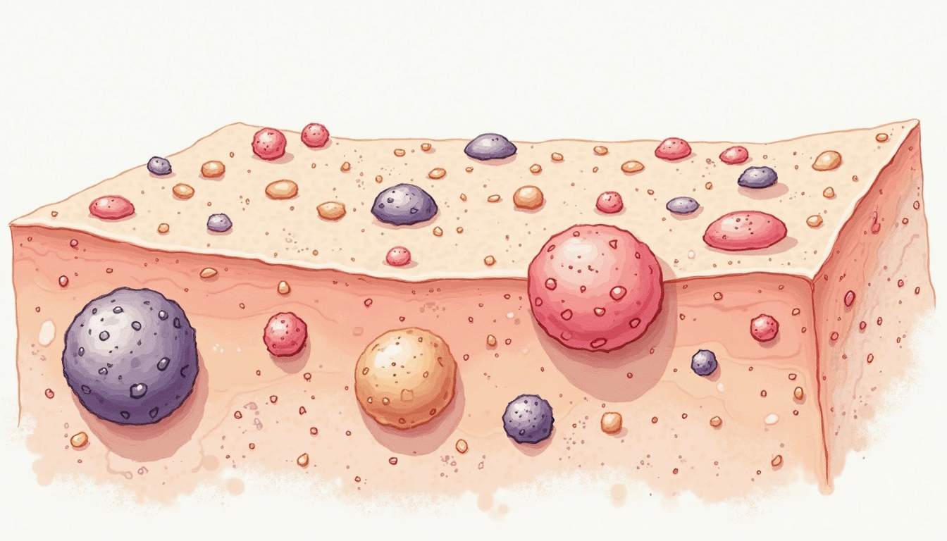
A nevus, commonly referred to as a mole, is a benign growth on the skin that arises from the proliferation of melanocytes, the cells responsible for producing the pigment melanin. Nevi can vary significantly in size, shape, and color, and they can appear anywhere on the body. While most nevi are harmless, some can develop into melanoma, a serious form of skin cancer, making it crucial for individuals to monitor their moles for any changes.
The study of nevi is an essential aspect of dermatology, as it encompasses various types, classifications, and potential health implications. Dermatologists often evaluate nevi during skin examinations, especially in patients with a history of skin cancer or those with numerous moles. Understanding the characteristics of nevi can aid in early detection and treatment of skin-related issues.
This glossary entry will explore the different types of nevi, their clinical significance, diagnostic criteria, treatment options, and preventive measures to maintain skin health. By comprehensively understanding nevi, individuals can better navigate their dermatological health and make informed decisions regarding their skin care.
Types of Nevi
Congenital Nevi
Congenital nevi are moles that are present at birth or develop shortly thereafter. They can vary in size from small to large and are classified based on their size and potential risk factors. Small congenital nevi are typically less than 1.5 cm in diameter, while large congenital nevi can exceed this measurement and may cover significant areas of the skin.
Large congenital nevi have a higher risk of developing into melanoma, particularly if they are larger than 20 cm. These nevi often require careful monitoring and, in some cases, surgical intervention to reduce the risk of malignancy. Congenital nevi can also be associated with other conditions, such as neurocutaneous melanosis, which involves the presence of melanocytic lesions in the central nervous system.
Acquired Nevi
Acquired nevi develop over time and are typically the result of sun exposure or other environmental factors. These moles usually appear during childhood or adolescence and can continue to change throughout a person's life. Acquired nevi are generally classified into several categories, including junctional nevi, compound nevi, and dermal nevi.
Junctional nevi are flat and located at the junction of the epidermis and dermis, while compound nevi are raised and consist of both epidermal and dermal components. Dermal nevi are entirely within the dermis and are usually raised and flesh-colored. Understanding the differences between these types of acquired nevi is essential for dermatologists when assessing potential risks and determining appropriate treatment options.
Other Types of Nevi
In addition to congenital and acquired nevi, there are several other specialized types of nevi that dermatologists may encounter. These include atypical nevi, also known as dysplastic nevi, which exhibit irregular features and may be larger than typical moles. Atypical nevi are often associated with an increased risk of melanoma and require careful monitoring.
Another type is the blue nevus, which is characterized by its blue or bluish-black color due to the depth of melanocytes within the dermis. While blue nevi are generally benign, they can sometimes be confused with melanoma, necessitating thorough evaluation by a dermatologist.
Clinical Significance of Nevi
Risk Factors for Melanoma
While most nevi are benign, certain characteristics can indicate an increased risk of melanoma. Factors such as a family history of skin cancer, the presence of numerous moles (more than 50), and atypical nevi can elevate an individual's risk. Additionally, individuals with fair skin, light hair, and light eyes are at a higher risk for developing melanoma.
Monitoring nevi for changes in size, shape, color, or texture is crucial for early detection of potential malignancies. Dermatologists often recommend regular skin checks, particularly for individuals with multiple risk factors. The ABCDE rule is a common guideline used to assess moles for signs of melanoma:
- A: Asymmetry - One half of the mole does not match the other half.
- B: Border - The edges are irregular, ragged, or blurred.
- C: Color - The color is not uniform and may include shades of brown, black, or tan.
- D: Diameter - The mole is larger than 6 mm (about the size of a pencil eraser).
- E: Evolving - The mole is changing in size, shape, or color.
Diagnosis of Nevi
Diagnosis of nevi typically involves a thorough clinical examination by a dermatologist. During this examination, the dermatologist will assess the characteristics of the mole, including its size, shape, color, and texture. If there are any concerns about the mole's appearance, a biopsy may be performed to obtain a sample of the tissue for further analysis.
Dermatologists may use dermatoscopes, specialized instruments that allow for magnified visualization of the skin, to enhance the examination of nevi. This tool can help identify subtle features that may indicate malignancy. In some cases, digital imaging may also be employed to monitor changes in moles over time.
Treatment Options for Nevi
Observation
For most benign nevi, the primary approach is observation. Regular monitoring allows dermatologists to track any changes in the mole's appearance. Patients are often advised to perform self-examinations and report any changes to their dermatologist during routine check-ups.
In cases where a nevus is deemed atypical or shows concerning changes, dermatologists may recommend a biopsy to determine whether it is benign or malignant. If the biopsy reveals melanoma, further treatment options will be discussed based on the stage of the cancer.
Surgical Removal
Surgical removal is a common treatment option for nevi that are suspected to be malignant or for those that cause cosmetic concerns. The procedure typically involves excising the mole along with a margin of surrounding skin to ensure complete removal of any potentially cancerous cells.
In some cases, dermatologists may opt for a shave excision, where the mole is shaved off the surface of the skin. This method is often used for raised nevi and may result in less scarring. However, it is essential to ensure that the entire mole is removed to prevent recurrence.
Preventive Measures
Sun Protection
One of the most effective ways to prevent the development of new nevi and reduce the risk of melanoma is through sun protection. Wearing broad-spectrum sunscreen with an SPF of 30 or higher is crucial when spending time outdoors. Sunscreen should be applied generously and reapplied every two hours, especially after swimming or sweating.
In addition to sunscreen, wearing protective clothing, such as long sleeves and wide-brimmed hats, can provide additional defense against harmful UV radiation. Seeking shade during peak sun hours, typically between 10 a.m. and 4 p.m., is also recommended to minimize sun exposure.
Regular Skin Examinations
Regular skin examinations by a dermatologist are vital for early detection of any changes in nevi. Individuals with a history of skin cancer or those with numerous moles should schedule annual check-ups or more frequent visits as recommended by their dermatologist. Self-examinations can also empower individuals to monitor their skin health and detect any concerning changes.
Education about the signs of melanoma and the importance of early detection can significantly impact outcomes. Understanding the ABCDE rule and being proactive about skin health can lead to timely interventions and better prognoses.
Conclusion
Nevi are common skin growths that can vary widely in appearance and significance. While most nevi are benign, understanding their characteristics and monitoring for changes is crucial for maintaining skin health and preventing melanoma. Dermatologists play a key role in evaluating, diagnosing, and treating nevi, providing essential care to patients at risk.
By prioritizing sun protection, regular skin examinations, and education about skin health, individuals can take proactive steps to manage their nevi and reduce their risk of skin cancer. Awareness and vigilance are essential components of dermatological care, ensuring that individuals can enjoy healthy skin throughout their lives.
Visit Our Offices
Services:
- • Medical Dermatology
- • Surgical Dermatology
- • Laser Treatments
- • Cosmetic Dermatology
Services:
- • Medical Dermatology
- • Surgical Dermatology
- • Laser Treatments
- • Cosmetic Dermatology
Visit Our Offices
Services:
- • Medical Dermatology
- • Surgical Dermatology
- • Laser Treatments
- • Cosmetic Dermatology
Services:
- • Medical Dermatology
- • Surgical Dermatology
- • Laser Treatments
- • Cosmetic Dermatology