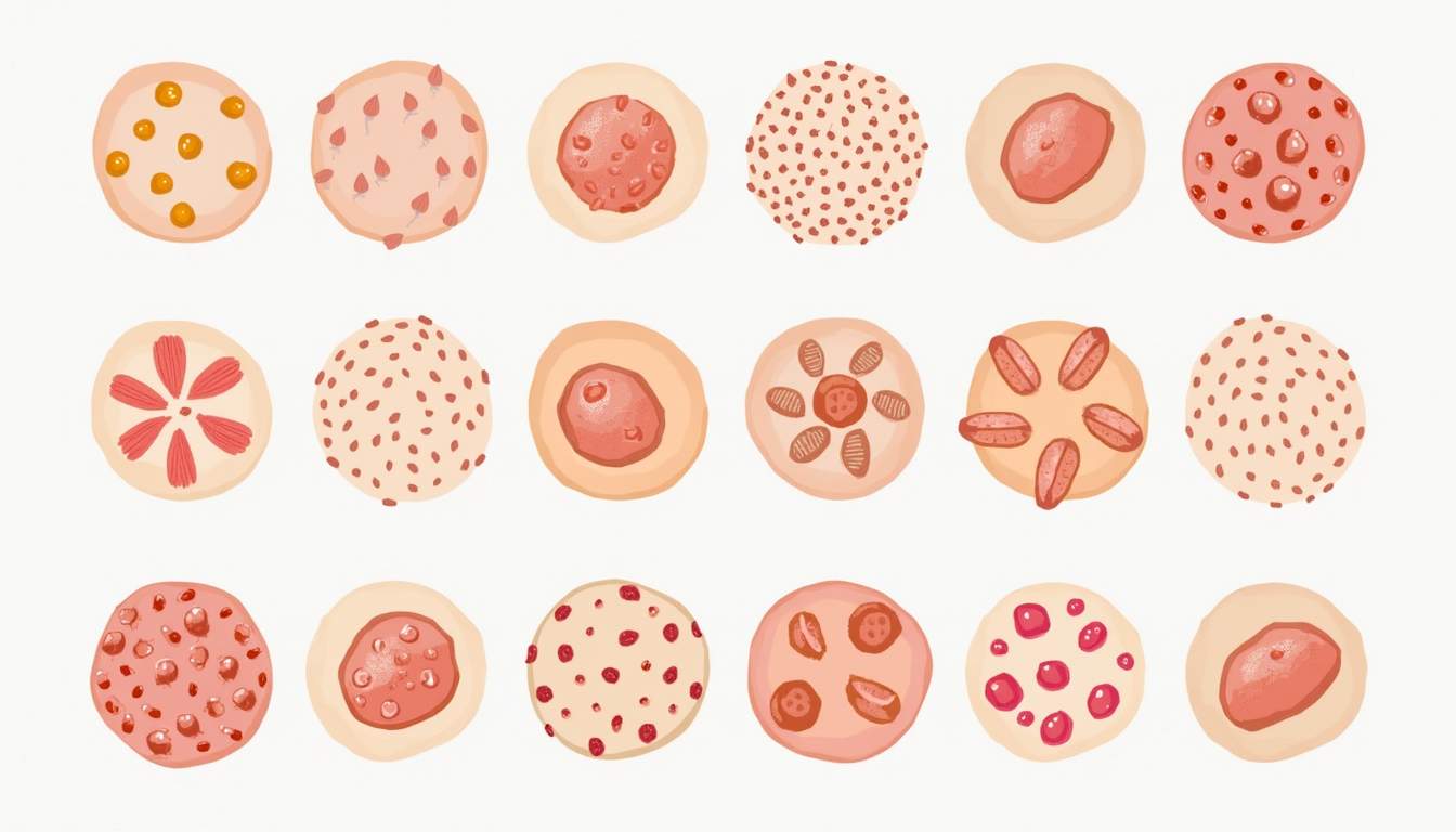
Introduction to Keratosis
Keratosis is a term used in dermatology to describe a variety of skin conditions characterized by the proliferation of keratinocytes, the predominant cell type in the outermost layer of the skin, known as the epidermis. These conditions can manifest in various forms, including but not limited to actinic keratosis, seborrheic keratosis, and keratosis pilaris. Each type of keratosis has distinct causes, symptoms, and treatment options, making it essential for both patients and healthcare providers to understand the nuances of these conditions.
The term 'keratosis' itself derives from the Greek word 'kerato,' meaning horn, which reflects the thickened, horn-like appearance of the affected skin. While keratosis is often benign, some forms, particularly actinic keratosis, can be precursors to skin cancer, emphasizing the importance of early detection and management.
In this glossary article, we will explore the various types of keratosis, their clinical features, underlying causes, diagnostic methods, and treatment approaches. This comprehensive overview aims to equip readers with a thorough understanding of keratosis within the broader context of dermatological health.
Types of Keratosis
Actinic Keratosis
Actinic keratosis (AK) is a common skin condition that arises due to prolonged exposure to ultraviolet (UV) radiation, primarily from the sun. It is characterized by rough, scaly patches on sun-exposed areas such as the face, ears, scalp, and backs of the hands. These lesions can vary in color from pink to red or brown and may feel dry or itchy. AK is considered a precancerous condition, as it can progress to squamous cell carcinoma if left untreated.
The risk factors for developing actinic keratosis include fair skin, a history of sunburns, older age, and a weakened immune system. Regular skin examinations by a dermatologist are crucial for early detection, especially for individuals at higher risk. Treatment options for AK may include topical therapies, cryotherapy, photodynamic therapy, and in some cases, surgical excision.
Seborrheic Keratosis
Seborrheic keratosis (SK) is a benign skin growth that typically appears as a raised, wart-like lesion with a waxy or scaly surface. These growths can vary in color from light tan to black and are often found on the trunk, face, scalp, and neck. Unlike actinic keratosis, seborrheic keratosis is not caused by sun exposure and is more common in older adults. The exact cause of SK is unknown, but genetic factors may play a role.
While seborrheic keratosis is harmless and does not require treatment, patients may choose to have them removed for cosmetic reasons or if they become irritated. Removal methods include cryotherapy, curettage, and laser therapy. It is essential to differentiate SK from other skin lesions, such as melanoma, which can be malignant. Therefore, any new or changing skin growth should be evaluated by a dermatologist.
Keratosis Pilaris
Keratosis pilaris (KP), often referred to as "chicken skin," is a common and harmless condition characterized by small, rough bumps on the skin, primarily on the upper arms, thighs, cheeks, and buttocks. These bumps are caused by the buildup of keratin, a protein that protects the skin, which clogs hair follicles. Keratosis pilaris is more prevalent in individuals with dry skin and can worsen in winter months or with certain skin conditions.
Although keratosis pilaris is not harmful and does not require treatment, many individuals seek remedies for cosmetic reasons. Moisturizers, exfoliating creams containing alpha-hydroxy acids, and retinoids can help improve the appearance of the skin. It's important to note that KP is often a chronic condition that may persist into adulthood, but it typically improves with time.
Causes of Keratosis
The causes of keratosis vary depending on the specific type. For instance, actinic keratosis is primarily caused by UV radiation exposure, which damages the DNA of skin cells and leads to abnormal growth. Factors such as geographic location, outdoor occupation, and personal habits like tanning can significantly increase the risk of developing AK.
Seborrheic keratosis, on the other hand, is believed to have a genetic component, as it often runs in families. The exact mechanisms behind its formation are still under investigation, but it is not linked to environmental factors like sun exposure. Keratosis pilaris is thought to be related to genetic predisposition and is often associated with other skin conditions, such as eczema or ichthyosis.
In summary, while some forms of keratosis are primarily influenced by environmental factors, others are more closely related to genetic predisposition. Understanding these underlying causes is crucial for effective prevention and management strategies.
Symptoms and Diagnosis
Symptoms of Keratosis
The symptoms of keratosis can vary widely depending on the type. Actinic keratosis typically presents as rough, scaly patches that may be itchy or tender. These lesions can range in size and may become more pronounced over time, often resembling a wart or dry skin. In contrast, seborrheic keratosis is characterized by raised, wart-like growths that can be smooth or scaly and may appear in clusters.
Keratosis pilaris presents as small, red or white bumps that feel rough to the touch. These bumps are often more noticeable on dry skin and may be accompanied by redness or inflammation. Importantly, keratosis pilaris does not cause pain or itching, making it more of a cosmetic concern than a medical one.
Diagnosis of Keratosis
Diagnosis of keratosis typically involves a thorough clinical examination by a dermatologist, who will assess the appearance and distribution of the lesions. In some cases, a skin biopsy may be performed to rule out other conditions, especially when there is uncertainty about the diagnosis. A biopsy involves removing a small sample of skin for laboratory analysis, which can provide definitive information about the type of keratosis and any potential malignancy.
Dermatologists may also utilize dermatoscopy, a non-invasive imaging technique that allows for a detailed examination of the skin's surface and subsurface structures. This method can aid in distinguishing between benign keratosis and more serious skin conditions, such as melanoma or basal cell carcinoma.
Treatment Options for Keratosis
Actinic Keratosis Treatments
Treatment for actinic keratosis is essential due to its potential to progress to skin cancer. Various treatment modalities are available, including topical therapies such as 5-fluorouracil (5-FU) and imiquimod, which work by inducing an inflammatory response that helps eliminate abnormal cells. Cryotherapy, which involves freezing the lesions with liquid nitrogen, is another common treatment option that can effectively remove AK lesions.
Photodynamic therapy (PDT) is a newer approach that combines a photosensitizing agent with light exposure to destroy abnormal cells. This method is particularly beneficial for patients with multiple actinic keratoses. In some cases, surgical excision may be necessary for larger or more persistent lesions.
Seborrheic Keratosis Treatments
For seborrheic keratosis, treatment is generally not required unless the lesions become bothersome or cosmetically undesirable. In such cases, dermatologists may recommend cryotherapy, curettage (scraping the lesion off), or laser therapy to remove the growths. These procedures are typically quick and can be performed in an outpatient setting.
It is important to note that while treatment can effectively remove seborrheic keratosis, new lesions may continue to develop over time, necessitating ongoing monitoring and management.
Keratosis Pilaris Treatments
Management of keratosis pilaris focuses on improving the skin's appearance and texture. Regular use of moisturizers can help alleviate dryness and reduce the rough texture associated with KP. Exfoliating agents, such as alpha-hydroxy acids (AHAs) or beta-hydroxy acids (BHAs), can help to remove dead skin cells and prevent follicle clogging. Retinoids may also be prescribed to promote cell turnover and improve skin texture.
While there is no definitive cure for keratosis pilaris, consistent skincare routines can significantly improve the condition. Patients are encouraged to be patient, as results may take time, and the condition may fluctuate with seasonal changes.
Prevention of Keratosis
Preventing keratosis, particularly actinic keratosis, primarily involves protecting the skin from UV radiation. This can be achieved through various strategies, including wearing broad-spectrum sunscreen with an SPF of 30 or higher, seeking shade during peak sun hours, and wearing protective clothing such as hats and long sleeves. Regular skin checks by a dermatologist can also aid in early detection and management of any suspicious lesions.
For seborrheic keratosis and keratosis pilaris, while prevention is less straightforward due to their genetic components, maintaining a healthy skincare routine can help manage symptoms. Regular moisturizing and gentle exfoliation can mitigate the appearance of keratosis pilaris, while monitoring for changes in seborrheic keratosis can ensure timely intervention if necessary.
Conclusion
Keratosis encompasses a range of skin conditions that, while often benign, require attention and understanding to ensure proper management and prevention of potential complications. From actinic keratosis, which poses a risk for skin cancer, to the more cosmetic concerns of seborrheic keratosis and keratosis pilaris, each type presents unique challenges and treatment options.
By fostering awareness and encouraging regular dermatological evaluations, individuals can take proactive steps in managing their skin health. Understanding the causes, symptoms, and treatment options for keratosis is crucial for both patients and healthcare providers, ultimately leading to better outcomes and enhanced quality of life.
Visit Our Offices
Services:
- • Medical Dermatology
- • Surgical Dermatology
- • Laser Treatments
- • Cosmetic Dermatology
Services:
- • Medical Dermatology
- • Surgical Dermatology
- • Laser Treatments
- • Cosmetic Dermatology
Visit Our Offices
Services:
- • Medical Dermatology
- • Surgical Dermatology
- • Laser Treatments
- • Cosmetic Dermatology
Services:
- • Medical Dermatology
- • Surgical Dermatology
- • Laser Treatments
- • Cosmetic Dermatology