Introduction to Histopathology
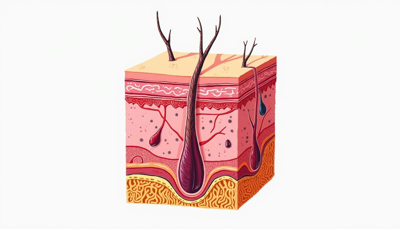
Histopathology is a branch of pathology that focuses on the microscopic examination of tissue samples to identify diseases. In dermatology, histopathology plays a crucial role in diagnosing skin disorders, including inflammatory conditions, infections, and neoplasms. By analyzing skin biopsies, dermatopathologists can provide essential insights into the underlying pathophysiology of various dermatological conditions.
The process of histopathological examination involves several steps, including tissue fixation, embedding, sectioning, staining, and microscopic evaluation. Each step is critical to ensure that the cellular architecture and any pathological changes are preserved and accurately represented. This meticulous approach allows for the identification of specific histological features that are characteristic of different skin diseases.
Histopathology not only aids in diagnosis but also helps in determining the prognosis and guiding treatment decisions. Understanding the histopathological aspects of dermatological conditions is essential for dermatologists and other healthcare providers involved in the management of skin diseases.
Importance of Histopathology in Dermatology
Histopathology is indispensable in dermatology for several reasons. Firstly, it provides a definitive diagnosis that can often be challenging to achieve based solely on clinical examination. Many skin conditions can present with similar clinical features, making it essential to obtain a tissue sample for accurate diagnosis. For instance, distinguishing between different types of dermatitis, psoriasis, and skin cancers often requires histopathological analysis.
Secondly, histopathology can reveal the extent of disease involvement. For example, in cases of melanoma, histopathological evaluation can determine the depth of invasion, which is crucial for staging the cancer and planning appropriate treatment. Additionally, histopathological findings can inform the likelihood of recurrence and metastasis, thereby influencing patient management strategies.
Moreover, histopathology contributes to the understanding of disease mechanisms. By examining the cellular and molecular changes in skin lesions, researchers and clinicians can gain insights into the pathogenesis of various skin disorders. This knowledge can lead to the development of targeted therapies and improved patient outcomes.
Key Techniques in Histopathology
Tissue Fixation
Tissue fixation is the first and one of the most critical steps in histopathology. It involves preserving the tissue sample to prevent autolysis and decay. Common fixatives include formaldehyde, which cross-links proteins and stabilizes cellular structures, and other agents like glutaraldehyde and alcohol. The choice of fixative can impact the quality of the histological analysis, as different fixatives preserve different cellular components.
The fixation process typically occurs shortly after the tissue is obtained, often within minutes, to ensure optimal preservation. The duration and conditions of fixation can vary depending on the type of tissue and the subsequent analysis planned. Proper fixation is essential for achieving high-quality histological sections that accurately represent the tissue architecture.
Embedding and Sectioning
Once the tissue is fixed, it is embedded in a medium, usually paraffin wax, to provide support for thin sectioning. The embedding process involves dehydrating the tissue and infiltrating it with the embedding medium. After embedding, the tissue block is cooled and solidified, allowing for precise cutting into thin sections, typically 4-5 micrometers thick.
Sectioning is performed using a microtome, a specialized instrument that allows for the creation of uniform tissue slices. The quality of the sections is critical, as uneven or thick sections can obscure histological details. Once sectioned, the tissue samples are placed on glass slides, ready for staining and microscopic examination.
Staining Techniques
Staining is a vital step in histopathology that enhances the visibility of cellular structures and components within the tissue. The most commonly used staining technique is Hematoxylin and Eosin (H&E) staining, which provides a general overview of tissue morphology. Hematoxylin stains cell nuclei blue, while eosin stains the cytoplasm and extracellular matrix pink, allowing for the differentiation of various cell types and structures.
In addition to H&E, various special stains and immunohistochemical techniques are employed to highlight specific cellular components or pathological changes. For example, immunohistochemistry uses antibodies to detect specific antigens in tissue sections, aiding in the diagnosis of conditions such as melanoma, where specific markers can indicate tumor type and behavior.
Common Dermatological Conditions Diagnosed by Histopathology
Inflammatory Skin Disorders
Histopathology is instrumental in diagnosing inflammatory skin disorders such as eczema, psoriasis, and dermatitis. In these conditions, histological examination typically reveals specific patterns of inflammation, including the presence of lymphocytes, eosinophils, and other immune cells. For instance, psoriasis is characterized by hyperkeratosis, acanthosis, and a dense infiltrate of T-lymphocytes in the dermis, which can be identified through histopathological analysis.
In cases of eczema, histopathology may show spongiosis, which is the accumulation of intercellular fluid within the epidermis, along with a mixed inflammatory infiltrate. These findings help differentiate eczema from other skin conditions that may present similarly, thereby guiding appropriate treatment strategies.
Infectious Skin Diseases
Histopathology is also crucial for diagnosing infectious skin diseases, including bacterial, viral, and fungal infections. For example, in cases of herpes simplex virus infection, histopathological examination may reveal multinucleated giant cells and intranuclear inclusions, which are characteristic of viral infections. Similarly, fungal infections such as dermatophytes can be identified through special staining techniques that highlight fungal hyphae and spores within the tissue.
Furthermore, histopathological analysis can help assess the extent of infection and the host's immune response, providing valuable information for treatment decisions. In cases of cutaneous abscesses or cellulitis, histopathology can reveal the presence of neutrophils and necrotic tissue, indicating an acute inflammatory response to infection.
Neoplastic Skin Conditions
Histopathology is essential for the diagnosis and classification of neoplastic skin conditions, including benign tumors such as seborrheic keratosis and malignant tumors like basal cell carcinoma and melanoma. The histological features of these tumors can vary significantly, and accurate identification is crucial for determining the appropriate management approach.
For instance, basal cell carcinoma typically exhibits nests of basaloid cells with peripheral palisading, while melanoma is characterized by atypical melanocytes and a lack of organized architecture. Histopathological examination allows for the assessment of tumor depth, mitotic activity, and other features that can influence prognosis and treatment options.
Challenges in Histopathological Diagnosis
Despite the advancements in histopathology, several challenges remain in the accurate diagnosis of dermatological conditions. One significant challenge is the potential for interobserver variability, where different pathologists may interpret the same histological features differently. This variability can be influenced by factors such as experience, training, and the complexity of the case.
Additionally, some skin conditions may exhibit overlapping histological features, making it challenging to arrive at a definitive diagnosis. For example, differentiating between various types of dermatitis or distinguishing between benign and malignant lesions can be particularly difficult. In such cases, additional diagnostic tools, such as molecular techniques or clinical correlation, may be necessary to achieve an accurate diagnosis.
Future Directions in Histopathology
The field of histopathology is continually evolving, with advancements in technology and techniques enhancing diagnostic capabilities. One promising area of development is the integration of digital pathology, which involves the use of digital imaging and artificial intelligence to assist in the analysis of histological slides. Digital pathology allows for more efficient storage, sharing, and analysis of histological data, potentially improving diagnostic accuracy and workflow efficiency.
Moreover, the incorporation of molecular techniques, such as next-generation sequencing and genomic profiling, is paving the way for personalized medicine in dermatology. These advancements enable the identification of specific genetic mutations and alterations associated with various skin conditions, leading to more targeted and effective treatment options.
As research continues to uncover the complexities of skin diseases, the role of histopathology will remain vital in bridging the gap between clinical practice and scientific understanding, ultimately improving patient care in dermatology.
Conclusion
In summary, histopathology is a cornerstone of dermatological practice, providing invaluable insights into the diagnosis, prognosis, and management of skin diseases. Through meticulous examination of tissue samples, dermatopathologists can identify a wide range of conditions, from inflammatory disorders to neoplastic lesions. The integration of advanced techniques and technologies continues to enhance the field, ensuring that histopathology will remain an essential component of dermatological care for years to come.
Visit Our Offices
Services:
- • Medical Dermatology
- • Surgical Dermatology
- • Laser Treatments
- • Cosmetic Dermatology
Services:
- • Medical Dermatology
- • Surgical Dermatology
- • Laser Treatments
- • Cosmetic Dermatology
Visit Our Offices
Services:
- • Medical Dermatology
- • Surgical Dermatology
- • Laser Treatments
- • Cosmetic Dermatology
Services:
- • Medical Dermatology
- • Surgical Dermatology
- • Laser Treatments
- • Cosmetic Dermatology