Introduction to Biopsy Techniques
A biopsy is a medical procedure that involves the removal of a small sample of tissue for examination under a microscope. In dermatology, biopsies are crucial for diagnosing skin conditions, including infections, inflammatory diseases, and skin cancers. The choice of biopsy technique depends on various factors, including the type of lesion, its location, and the clinical suspicion of the underlying pathology.
Understanding the different biopsy techniques available in dermatology is essential for both clinicians and patients. Each technique has its indications, advantages, and disadvantages. This glossary entry aims to provide a comprehensive overview of the various biopsy methods, their procedural steps, and the implications of the results obtained from these procedures.
In dermatology, the most common biopsy techniques include punch biopsy, shave biopsy, excisional biopsy, and incisional biopsy. Each of these methods serves a specific purpose and is chosen based on the characteristics of the skin lesion being evaluated.
Punch Biopsy
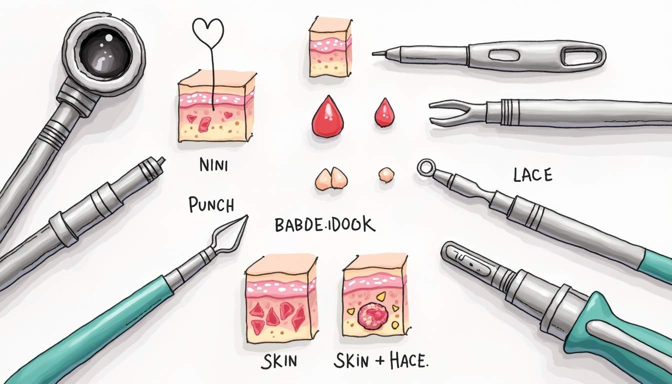
A punch biopsy is a technique that uses a circular blade to remove a cylindrical core of skin, including the epidermis, dermis, and sometimes subcutaneous tissue. This method is particularly useful for diagnosing conditions such as psoriasis, dermatitis, and skin cancers. The size of the punch can vary, typically ranging from 2 mm to 8 mm in diameter, depending on the lesion's size and the depth of tissue required for diagnosis.
The procedure begins with the application of a local anesthetic to minimize discomfort. Once the area is numb, the punch tool is pressed down onto the skin, rotating it to cut through the layers of tissue. After the core is removed, the wound is usually closed with sutures, although smaller biopsies may heal well without stitches. The tissue sample is then sent to a laboratory for histopathological examination.
One of the advantages of punch biopsies is that they provide a full-thickness sample of the skin, allowing for a more accurate diagnosis. Additionally, they are relatively quick to perform and can be done in an outpatient setting. However, the size of the punch may limit the amount of tissue available for certain types of analyses, and there is a risk of scarring or infection at the biopsy site.
Indications for Punch Biopsy
Punch biopsies are indicated for a variety of dermatological conditions, including:
- Skin cancers, such as melanoma and basal cell carcinoma
- Inflammatory skin diseases, including eczema and psoriasis
- Vesicular lesions, such as those seen in herpes simplex virus infections
- Uncertain rashes where a definitive diagnosis is needed
By providing a full-thickness sample, punch biopsies can help differentiate between various skin conditions that may present similarly on the surface. This is particularly important in cases where the clinical appearance is ambiguous, and a precise diagnosis is necessary for effective treatment.
Shave Biopsy
A shave biopsy is a technique that involves the removal of a superficial layer of skin using a surgical blade. This method is commonly used for lesions that are raised above the surrounding skin, such as seborrheic keratoses, warts, and some skin cancers. The shave technique allows for the removal of the lesion while preserving the surrounding healthy skin, which can be beneficial for cosmetic reasons.
During the procedure, a local anesthetic is administered to the area around the lesion. The clinician then uses a scalpel or a specialized shave tool to carefully shave off the lesion at the level of the surrounding skin. The resulting wound is usually left to heal without sutures, as it is typically small and will close on its own. The excised tissue is sent for pathological evaluation to determine the nature of the lesion.
Shave biopsies are advantageous due to their simplicity and speed. They can often be performed in a matter of minutes and do not require extensive recovery time. However, one limitation is that shave biopsies may not provide a full-thickness sample of the skin, which can be a concern when evaluating deeper lesions or when a complete diagnosis is required.
Indications for Shave Biopsy
Shave biopsies are particularly useful for:
- Benign lesions, such as dermatofibromas and seborrheic keratoses
- Superficial skin cancers, where complete excision is not necessary
- Lesions that are easily accessible and do not require deep tissue sampling
By utilizing the shave biopsy technique, dermatologists can efficiently diagnose and treat superficial skin lesions while minimizing patient discomfort and recovery time. However, it is essential to recognize that this method may not be suitable for all types of lesions, particularly those with a high suspicion of malignancy.
Excisional Biopsy
An excisional biopsy is a surgical procedure that involves the complete removal of a skin lesion along with a margin of surrounding healthy tissue. This technique is often employed when there is a high suspicion of malignancy, as it allows for both diagnosis and treatment in a single procedure. Excisional biopsies are commonly performed on larger lesions or those located in areas where a clear margin is necessary for accurate diagnosis.
The procedure typically begins with the administration of local anesthesia to numb the area. The clinician then uses a scalpel to excise the lesion, ensuring that a margin of healthy tissue is included. The wound is usually closed with sutures, and the excised tissue is sent for pathological examination. The histopathological analysis will provide information on the nature of the lesion and whether further treatment is necessary.
One of the primary advantages of excisional biopsies is that they provide a comprehensive sample for analysis, allowing for accurate diagnosis and staging of skin cancers. Additionally, by removing the entire lesion, excisional biopsies can prevent the need for subsequent procedures. However, this technique may result in larger scars compared to other biopsy methods, and there is a risk of complications such as infection or delayed healing.
Indications for Excisional Biopsy
Excisional biopsies are indicated for:
- Suspicious moles or lesions with features suggestive of melanoma
- Large or deep skin cancers requiring complete removal
- Lesions that have not responded to other treatments
By employing excisional biopsy techniques, dermatologists can ensure that potentially malignant lesions are thoroughly evaluated and treated, reducing the risk of recurrence and improving patient outcomes. This method is particularly important in cases where early detection and intervention are critical.
Incisional Biopsy
An incisional biopsy is a technique that involves the removal of a portion of a larger lesion rather than the entire mass. This method is often used for lesions that are too large to be completely excised or when the clinical suspicion of malignancy is uncertain. Incisional biopsies allow for the collection of tissue for diagnostic purposes while preserving as much healthy tissue as possible.
The procedure begins similarly to other biopsy methods, with the administration of local anesthesia. The clinician then makes an incision through the skin to remove a representative portion of the lesion. The sample is sent for histopathological evaluation, which will provide information on the nature of the lesion and guide further management.
Incisional biopsies are advantageous in that they allow for the evaluation of larger lesions without the need for complete excision. This can be particularly useful in cases where the lesion is located in a cosmetically sensitive area or when the patient has other medical conditions that make extensive surgery risky. However, like excisional biopsies, there is a risk of complications such as infection and scarring.
Indications for Incisional Biopsy
Incisional biopsies are indicated for:
- Large tumors where complete excision is not feasible
- Lesions with uncertain diagnosis that require further evaluation
- Cases where preserving surrounding tissue is essential for cosmetic reasons
By utilizing incisional biopsy techniques, dermatologists can obtain necessary diagnostic information while minimizing the impact on the patient's appearance and overall health. This approach is particularly valuable in managing complex cases where a careful balance between diagnosis and treatment is required.
Conclusion
In summary, biopsy techniques in dermatology play a vital role in the diagnosis and management of various skin conditions. Understanding the different methods, including punch biopsy, shave biopsy, excisional biopsy, and incisional biopsy, is essential for both healthcare providers and patients. Each technique has its specific indications, advantages, and limitations, and the choice of method should be tailored to the individual patient's needs and the characteristics of the lesion being evaluated.
As dermatology continues to evolve, advancements in biopsy techniques and technologies will likely enhance diagnostic accuracy and patient outcomes. Ongoing education and training for dermatologists in these techniques are crucial to ensure that patients receive the best possible care. Ultimately, the goal of any biopsy procedure is to provide a definitive diagnosis that guides effective treatment and improves the patient's quality of life.
For patients undergoing a biopsy, understanding the procedure and its implications can help alleviate anxiety and foster a collaborative relationship with their healthcare provider. Open communication about the reasons for the biopsy, the expected outcomes, and the potential risks can empower patients to make informed decisions about their dermatological care.
Visit Our Offices
Services:
- • Medical Dermatology
- • Surgical Dermatology
- • Laser Treatments
- • Cosmetic Dermatology
Services:
- • Medical Dermatology
- • Surgical Dermatology
- • Laser Treatments
- • Cosmetic Dermatology
Visit Our Offices
Services:
- • Medical Dermatology
- • Surgical Dermatology
- • Laser Treatments
- • Cosmetic Dermatology
Services:
- • Medical Dermatology
- • Surgical Dermatology
- • Laser Treatments
- • Cosmetic Dermatology